AC joint dislocations
Acute care & follow-up treatment
An acromioclavicular joint(AC joint dislocation) is an injury to the small joint between the acromion(shoulder roof) and the outermost part of the clavicle(collarbone). This injury is usually caused by direct trauma to the shoulder from the front, as is often the case when falling off a bicycle, over the handlebars or while snowboarding.
OVERVIEW
Symptoms of an AC joint fusion
Typically, a swelling is visible directly above the acromioclavicular joint, as well as a higher collarbone compared to the opposite side. As the severity of the injury increases, the misalignment and dislocation of this joint is easier to recognize and causes increased discomfort , especially when working overhead or doing sports.
Diagnosis of an AC joint fusion
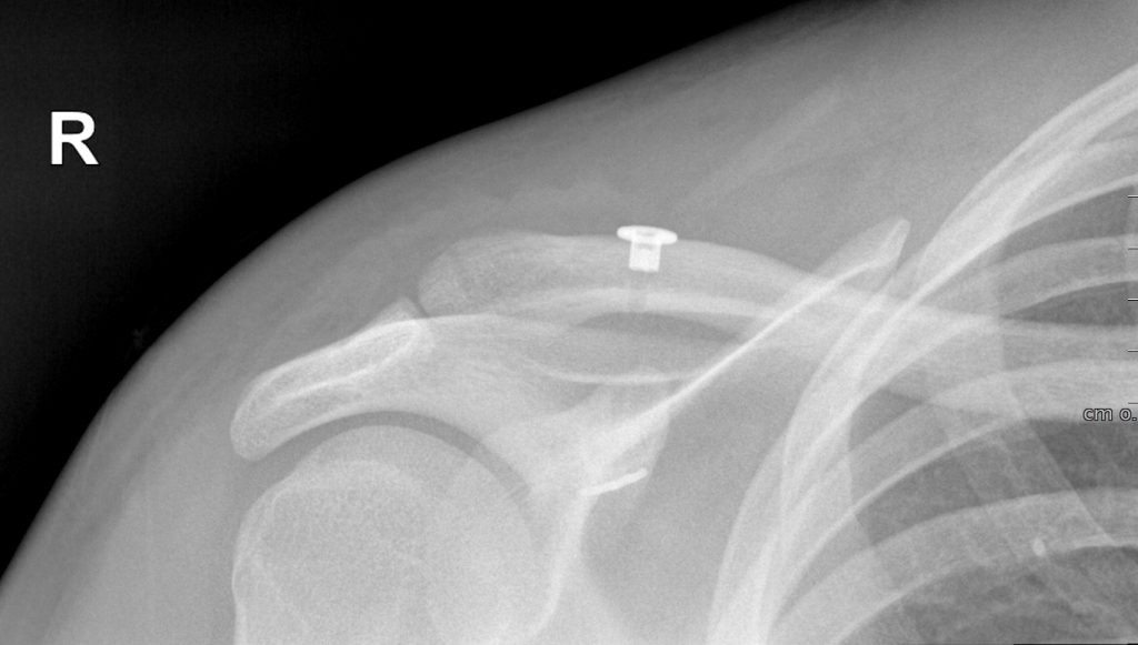
In addition to a clinical examination of the shoulder and a detailed medical history, specific x-rays will be taken to show the severity and extent of the accompanying injuries. This injury is classified according to Rockwood based on the position of the outer part of the clavicle. This classification helps us to derive the necessary therapy.
Classification of severity according to Rockwood I-VI
- Type I: Stretching/straining of the ligamentous apparatus
- Type II: Injury to the joint capsule of the acromioclavicular joint with minimal elevation of the clavicle
- Type III: Complete tear of the joint capsule and the ligamentous connection between the collarbone and the shoulder blade. The collarbone is raised by up to 100%
- Type IV: In addition to the vertical displacement of the clavicle, horizontal translation occurs. The collarbone is stuck in the trapezius muscle here.
- Type V: In addition to type III, there is a partial extensive detachment of the muscle insertions and thus a protrusion of the clavicle by more than 100%
- Type VI: Hacking of the clavicle under the coracoid process of the scapula. Very rare!
Treatment of an AC joint fusion
Treatment depends on the severity of the injury. Types I and II are treated conservatively with a shoulder bandage and subsequent physiotherapy.
Completely torn ligaments(type III) can be treated either conservatively or surgically. After conservative treatment, often only the protruding collarbone remains cosmetically unpleasant, but shoulder function is usually comparable with both treatment approaches. It is important to distinguish whether a type III injury involves only vertical or also horizontal instability, as surgery should be favored for more complex instabilities. Athletes (overhead sports: tennis, volleyball,…) with high loads on the shoulder also tend to benefit more from surgery.
Surgery is clearly superior to conservative therapy for types IV-VI
Surgery for an AC joint fusion
I perform the operation minimally invasively by means of arthroscopy (joint endoscopy). Here the collarbone is reattached to the shoulder blade with 2 suture systems and metal buttons. In addition, the AC joint is wrapped in 8 turns with a Fibretapem (artificial ligament) to also address horizontal instability. This is a very stable fixation, with only small surgical scars, without the need to remove a metal implant in the process. Primary surgery is only possible in the first 3 weeks after the accident, as otherwise the torn ligaments are already scarred without guaranteeing stability.
After 3 weeks, the biology must be additionally stimulated in order to achieve good long-term results. A tendon is therefore harvested from the thigh and used as an additional augmentation of the technique described above.